Computed Tomography (CT) scanners are often the first imaging technology many patients encounter when doctors suspect serious disease or injury. The machines use a narrow beam of X-rays processed by a computer to create slices of the body and assemble them into detailed 3D images.
Additionally, Revolution CT is able to complete the scan using up to 82% less ionizing radiation than traditional CT scanners, which is ideal in use with pediatric and oncology patients. The machine is also sensitive enough that it could obtain images using less contrast dye, which is welcome news for those who are sensitive to those chemicals.
The Revolution CT is able to get such clear images due to the camera’s motion correction, which is very similar to the image stabilizing technology found inside handheld point-and-shoot cameras. This allows the machine to correct for any fast movements and reduce noise in the picture, even when faced with rapid heartbeats. It is equipped with the fastest scintillator in the industry, capturing the images very quickly. Additionally, the machine’s collimator reduces the scatter of the beams, which helps resolve artifacts that occur during the imaging process.

Above: A high-definition musculoskeletal image of a foot and ankle reinforced with plates and screws.
In 2013, GE introduced a new, superfast scanner called Revolution CT that allowed doctors to routinely obtain clear images of the beating heart, lungs, liver and other organs.

[Above] An image of the abdomen and the aorta.
Starting in September 2014, the West Kendall Baptist Hospital in Florida became the first medical facility in the U.S. to use the machine. Its combination of low-dose exposure, organ-wide coverage and motion correction technology allows doctors to reduce radiation and still obtain high-resolution images of blood vessels, soft tissue, organs and bones.

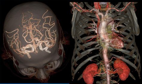
[Above] The whole aorta and kidneys.
The team at West Kendall Baptist Hospital recently completed the world’s first six-month clinical trial of the Revolution CT machine. Local doctors said they were able to diagnose even the most challenging cardiac patients with erratic or high heartbeats and reduce the radiation dose for pediatric patients.
“According to our physicians, patient feedback about their experience with the Revolution CT has been uniformly positive,” said West Kendall Baptist Hospital CEO Javier Hernández-Lichtl. “The advanced design definitely makes for a less intimidating, more comfortable patient experience, while yielding amazingly accurate and detailed images.”

A high-definition image of the skull and the Circle of Willis.
The Revolution CT was developed by scientists and engineers at GE Healthcare and GE Global Research, who were working closely with physicians in the field. “A core component of our strategy at GE Healthcare is to partner with customers to understand their clinical and operational needs, and in turn develop next-generation technology that deliver the necessary outcomes,” said Jeff Immelt, GE chairman and CEO, who came to West Kendall to see the results.
Take a look at some of the images obtained by the machine.

[Above] The Circle of Willis.

[Above] A high-definition image of the skull and the Circle of Willis.

[Above] The skull and carotid arteries.

[Above] An image of the abdomen and pelvis.

[Above] The rib cage, the heart and the chest cavity. The Revolution CT can image the heart in a single heartbeat.

[Above] An image of the human heart with stents typically used to treat narrow or weak arteries.

[Above] The chest cavity with a side view of the heart.

[Above] The pelvis and the aorta.

[Above] The whole aorta and kidneys.

A high-definition musculoskeletal image of a foot with a screw.


Above image: A high-definition image of the skull and the Circle of Willis, which supplies blood to the brain.
[All images credited to: GE Healthcare]

![FDA Forced to Retract Misleading Statements on Ivermectin’s COVID-19 Use After Legal Challenge [VIDEO]](https://i0.wp.com/austincountynewsonline.com/wp-content/uploads/2016/05/fda.jpg?resize=440,264)

:quality(75)/https://static.texastribune.org/media/files/4b25469bb109b715051f54458791074f/0303%20Panhandle%20Canadian%20JR%20TT%2025.jpg?resize=440,264 "First Human Case Of Bird Flu In Texas Detected After Contact With Infected Dairy Cattle")
